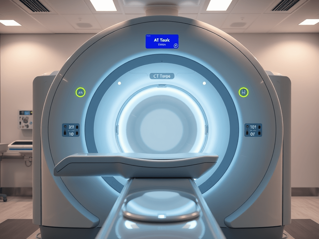
A CT perfusion study is a medical imaging technique that evaluates the blood flow to various tissues, particularly in the brain or heart. It uses computed tomography (CT) technology in conjunction with a contrast agent to visualize how blood is delivered to different areas over time.
In a CT perfusion study, a series of images is taken rapidly after the injection of contrast material, allowing the physician to assess parameters such as cerebral blood flow (CBF), cerebral blood volume (CBV), and mean transit time (MTT). This information is essential for diagnosing conditions like stroke, tumors, or other vascular diseases, as it helps identify areas of reduced blood supply or ischemia.
Here are some interesting facts about CT perfusion studies:
1. Rapid Imaging: CT perfusion studies can capture a series of images in just a few seconds, allowing for real-time assessment of blood flow dynamics in tissues.
2. Stroke Assessment: One of the primary uses of CT perfusion is in the evaluation of acute ischemic stroke. It helps to differentiate between viable brain tissue and areas that have already undergone irreversible damage.
3. Quantitative Analysis: The study provides quantitative data on blood flow parameters, such as cerebral blood flow (CBF), cerebral blood volume (CBV), and mean transit time (MTT), which can guide treatment decisions.
4. Contrast Agent: The procedure typically involves the use of a iodinated contrast agent, which enhances the visibility of blood vessels and perfusion patterns on the CT images.
5. Multi-Modal Approach: CT perfusion can be combined with other imaging techniques, such as CT angiography or MRI, to provide a comprehensive assessment of vascular and tissue health.
6. Non-Invasive: CT perfusion is a non-invasive procedure, making it a preferred choice for patients who cannot undergo more invasive tests.
7. Applications Beyond the Brain: Although commonly used for neurological assessments, CT perfusion studies can also be utilized for evaluating perfusion in other organs, such as the heart, liver, and kidneys.
8. Guiding Interventions: The data obtained from CT perfusion studies can help in planning surgical interventions, such as revascularization procedures in cases of blocked blood vessels.
9. Speed of Diagnosis: The quick nature of CT perfusion studies can lead to faster diagnosis and treatment, which is critical in time-sensitive conditions like stroke.
10. Research and Development: Ongoing research continues to refine the techniques and algorithms used in CT perfusion studies, enhancing their accuracy and applicability in clinical practice.
here is an additional 27 minutes of CT perfusion made easy, enjoy.

Leave a comment