Ever wonder why Dr. Haycock asks for the TEE kit when we have a full arrest coming into the emergency department? Let’s look into the simple yet valuable tool used during a code situation.
Why TEE for cardiopulmonary resuscitation
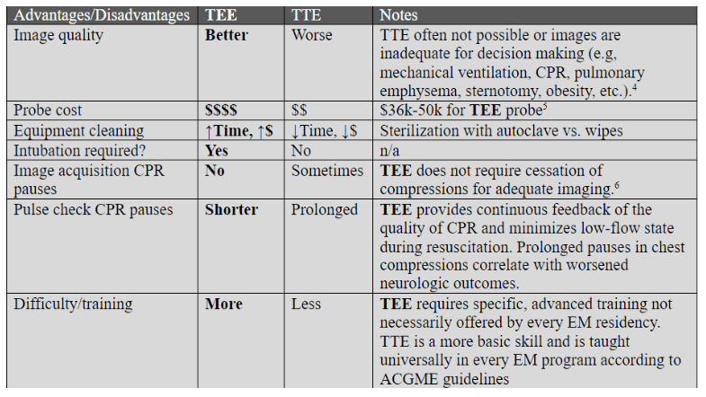
TEE facilitates continuous cardiac monitoring, aids in identifying reversible causes, optimizes chest compression techniques, can notably shorten CPR pauses, and helps differentiate between various cardiac activities. Lots of good stuff!
Difference between a TTE and a TEE
TTE involves placing the transducer on the chest wall, while TEE involves inserting a transducer into the esophagus.
Diving deeper into the mitral valve and PVF
While the thoracic pump theory suggests the heart is a passive conduit, evidence indicates that the mitral valve can fulfill a more active role, particularly when the heart retains some capacity for contraction. The movement of the mitral valve is crucial for maintaining sufficient blood flow during CPR, directly impacting the success of resuscitation efforts.
Since the PVF (pulmonary venous flow) constitutes the inflow of blood into the left atrium and the left ventricle, it should be considered alongside mitral valve position and transmitral flow. If the chest pump model holds true and the left heart functions merely as a conduit, mitral valve opening should occur with forward PVF (from the pulmonary vein into the left atrium) during chest compression.
Uenishi et al. stated that the pressure gradient during chest compression follows the order of left atrium > left ventricle > aorta, suggesting that the left atrium is the primary contributor to the flow generated in the early phase of compression.
Watch Brief intro to Resuscitative TEE | https://www.youtube.com/watch?v=gQw1aKYBdrA |

Leave a comment