Case study: Mr. J. is a 76 year old gentleman who came in with left sided weakness associated with dizziness and visual changes. He was recently started on an anti-amyloid therapies Lecanemab a few weeks back to help slow down his Alzheimer’s disease progression. He is AO x 3 at baseline. He is currently a GCS of 4-4-5. His LKWT was 2 hours ago, BP 178/105, BG 178, sinun rhythm on the monitor and is on baby ASA daily. Other pertinent history are HNT, DM2, and hyperlipidemia.
ARIA and Anti-Amyloid Therapy
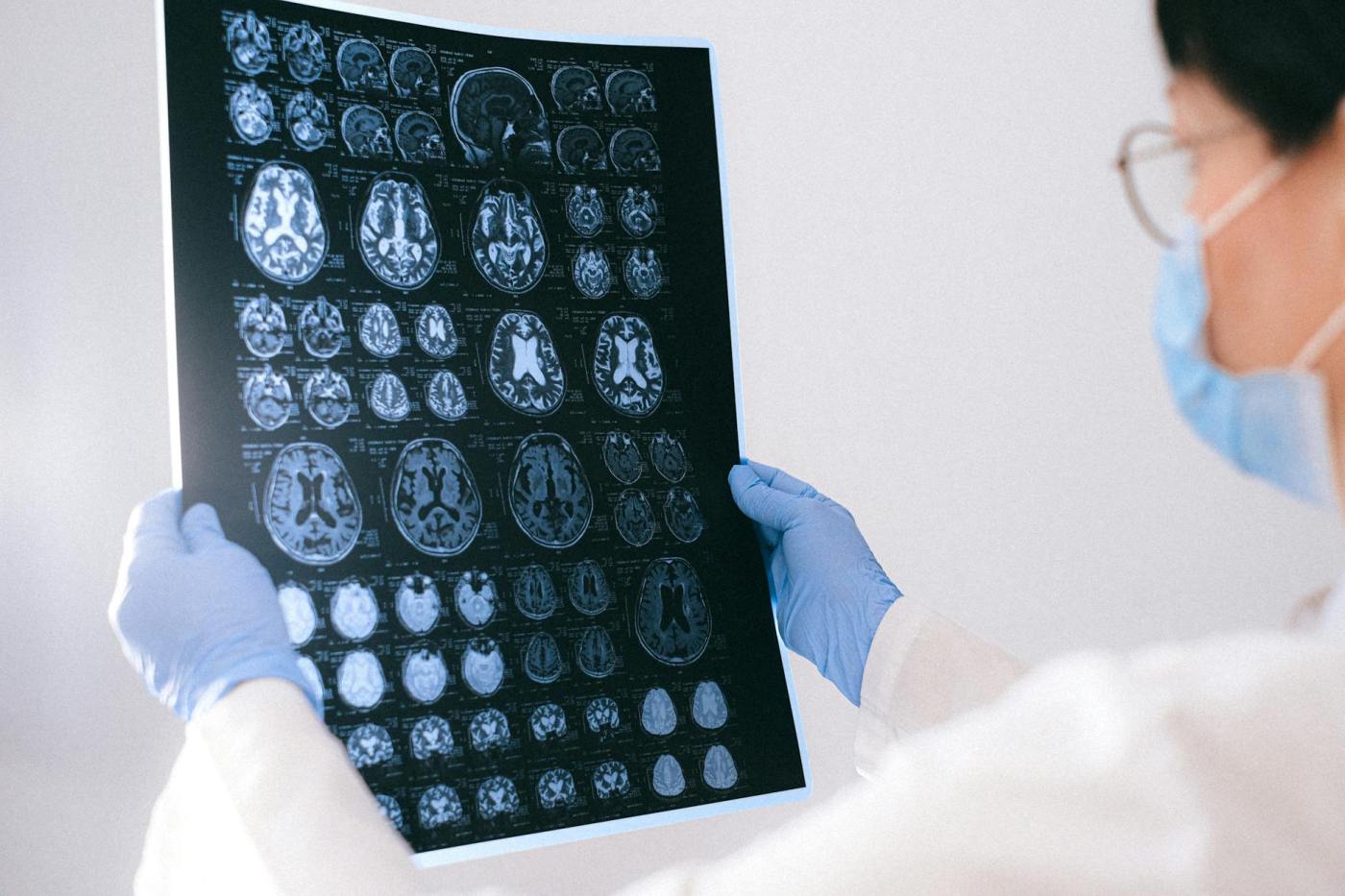
Anti-amyloid therapies, such as lecanemab and donanemab, help reduce amyloid plaques in the brain and can slow Alzheimer’s disease progression. However, they may cause ARIA, which appears as abnormalities on MRI scans. ARIA is a side effect seen in patients receiving monoclonal antibodies (MAb) targeting amyloid-beta (Aβ) in Alzheimer’s trials. While these treatments aim to clear amyloid plaques, they can also trigger inflammation and result in ARIA (Amyloid-related imaging abnormalities).
ARIA-E and ARIA-H
ARIA-E, or edema, means fluid buildup in the brain, while ARIA-H, or hemorrhage, involves tiny bleeds or iron deposits. Both types can be seen on MRI. Although ARIA-E and ARIA-H usually don’t show symptoms, some patients might have headaches, confusion, or other neurological issues. Occasionally, ARIA-H is linked to symptomatic intracerebral hemorrhage, and there have also been reports of ischemic strokes.
Underlying Pathophysiology
The exact reasons for ARIA are not completely clear, but it may be linked to more leaky blood vessels (increase permeability), inflammation, and how antibodies affect a brain condition called cerebral amyloid angiopathy (CAA). CAA occurs when amyloid builds up in brain blood vessels and is seen as a risk factor for ARIA.
Conclusion
In summary, ARIA is a common side effect of anti-amyloid immunotherapy and can sometimes resemble a stroke, causing neurological symptoms. It’s important to understand ARIA’s mechanisms, risk factors, and clinical signs to effectively manage patients using these treatments and to differentiate ARIA from other neurological issues. Amyloid-related imaging abnormalities (ARIA), especially ARIA-H (hemorrhage) and ARIA-E (edema), may pose risks with anti-amyloid therapy. Thrombolytic therapy, such as tissue plasminogen activator (t-PA) for stroke, could heighten the risk of ARIA-H in patients receiving anti-amyloid treatment. Thus, using thrombolytics in these patients should be carefully considered and may not be advisable.
We need to continue to consult our in house and on call neurologist for patient within the 4.5 hours of LKWT and make them aware of these two Anti-amyloid therapies in formulating our inclusion and exclusion for possible thrombolytic therapy.
Names, dates, and personal identifying details have been changed throughout this website to comply with the Health Insurance Portability and Accountability Act (HIPAA). **.

Leave a comment